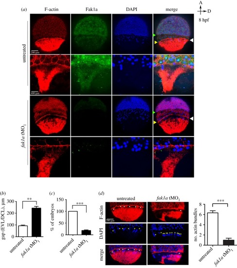
Loss of Fak1a perturbs the synchronized migration of enveloping and deep cell layers and the F-actin network. (a) Embryos untreated or injected with 5 ng of a fak1a translation blocking morpholino (tMO1) were fixed at 8 hpf and subjected to F-actin/DAPI staining and Fak1a immunohistochemistry. Embryos were examined and photographed for the whole embryo image (upper) or a region flanking the yolk syncytial at a higher magnification (lower) under confocal microscopy. Representative photographs of different channels and merged images are shown. White arrowheads point to actin rings. Yellow and green arrowheads point to the running fronts of the enveloping (EVL) and deep cell layers (DCL), respectively. (b) Graphic demonstration of the average gap between EVL and DCL in untreated and fak1a tMO1-treated embryos (n = 3; n = 30, **p < 0.01). (c) The EVL/DCL gap smaller than 100 µm were considered normal, and the percentages of normal embryos are presented (n = 3; n = 30, ***p < 0.001). (d) The actin bundles (marked by white asterisks) between the actin ring and vegetal actin cap were clearly reduced in fak1a morphants, and the numbers of actin bundles are quantified in the right panel (n = 3; n = 10, ***p < 0.001). Disorganized YSL nuclei (yellow asterisks) were also observed in fak1a morphants (see DAPI staining and merged images). As indicated by the arrows in the top right corner, all images have the animal pole (a) placed at the top and the dorsal to the right (d).
|
