FIGURE

Fig 2

ID
ZDB-FIG-200225-2
Publication
Belzunce et al., 2020 - The interplay of atoh1 genes in the lower rhombic lip during hindbrain morphogenesis
Other Figures
All Figure Page
Back to All Figure Page
Fig 2

Analysis of the <italic>atoh1a</italic> neuronal derivatives in Tg[atoh1a:GFP] embryos.

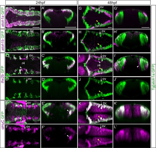
Tg[atoh1a:GFP] embryos at 24hpf and at 48hpf were assayed for atoh1a (A, G), atoh1b (B, H), lhx2b (C-D, I-J) in situ hybridization, and anti-HuC (E-F, K-L) staining. Dorsal views of confocal MIP from dorsal stacks (A-B) or ventral stacks (C-L) with anterior to the left. A’-L’) Reconstructed transverse sections of the dorsal views in (A-L) at the level indicated with the white arrow depicted in (A-L) corresponding to r4/r5. All embryos displayed the atoh1a-progenitors and derivatives in green. Note that atoh1b cells derive from atoh1a:GFP progenitors (B’, H’), as well as the lateral lhx2b neuronal domain (see white asterisks in C’, I-I’), whereas the medial lhx2b neuronal column in r4 is devoid of green staining (see white arrowhead in C, I-I’). See that differentiated neurons organize in arch-like structures (yellow arrowhead in G-L). ov, otic vesicle; SAG, statoacoustic ganglion; r, rhombomere. Scale bars correspond to 50 μm.

Expression Data
Genes:
Fish:
Anatomical Terms:
Stage Range: Prim-5 to Long-pec

Expression Detail
Antibody Labeling
Phenotype Data

Phenotype Detail
Acknowledgments
This image is the copyrighted work of the attributed author or publisher, and ZFIN has permission only to display this image to its users. Additional permissions should be obtained from the applicable author or publisher of the image. Full text @ PLoS One