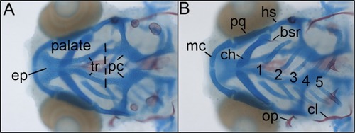
Wild‐type zebrafish craniofacial anatomy. Dorsal (A) and ventral (B) views of a 5 dpf whole mount Alcian Blue/Alizarin Red‐stained zebrafish. Our forward genetic screen identified EtOH‐sensitive mutants with qualitative alterations to the craniofacial skeleton. (A) The neurocranium consists of an anterior, neural crest‐derived palatal skeleton, composed of the trabeculae (tr) and ethmoid plate (ep). The palate connects posteriorly to the mesoderm‐derived parachordal cartilages (pc). (B) The viscerocranium consists of 7 segments. The first and second segments have distinct ventral and dorsal skeletal elements at this age. Meckel’s cartilage (mc) and the palatoquadrate (pq) reside ventrally and dorsally, respectively, in the first pharyngeal arch. Ventral and dorsal elements within the second pharyngeal arch are the ceratohyal cartilage (ch) with its associated bone the branchialstegal ray (bsr) and the hyosymplectic cartilage (hs) and opercle bone (op), respectively. The remaining 5 pharyngeal arches house ceratohyal cartilages 1 to 5 (numbered), with the fifth ceratohyal harboring the pharyngeal teeth. The mesoderm‐derived cleithrum (cl) resides just posterior to the pharyngeal arches. Modified from Swartz and colleagues (2014).
|
