
- Title
-
Novel ethanol-sensitive mutants identified in an F3 forward genetic screen
- Authors
- Swartz, M.E., Lovely, C.B., McCarthy, N., Kuka, T., Eberhart, J.K.
- Source
- Full text @ Alcoholism Clin. Exp. Res.
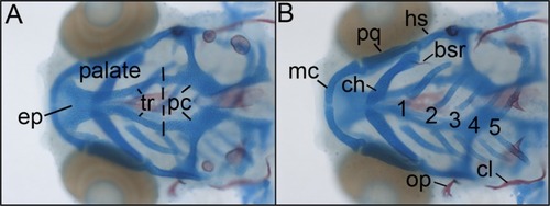
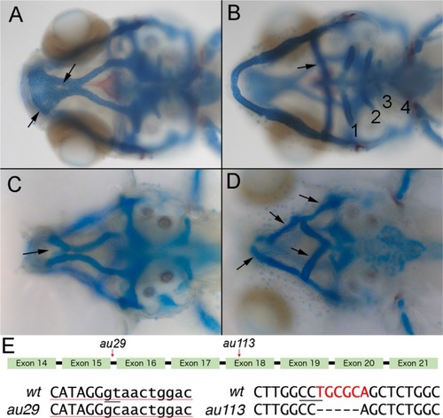
Wild‐type zebrafish craniofacial anatomy. Dorsal ( |
The PHENOTYPE:
|
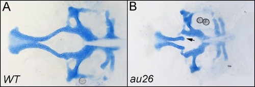
Lower jaw defects in PHENOTYPE:
|
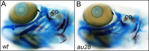
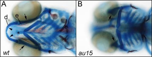
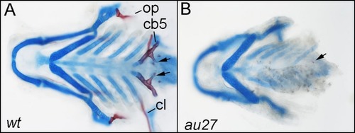
Hyomandibular defects in EtOH‐treated PHENOTYPE:
|
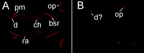
Bone loss in EtOH‐treated PHENOTYPE:
|
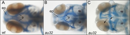
Bone loss in PHENOTYPE:
|
Phenotypic classes of PHENOTYPE:
|
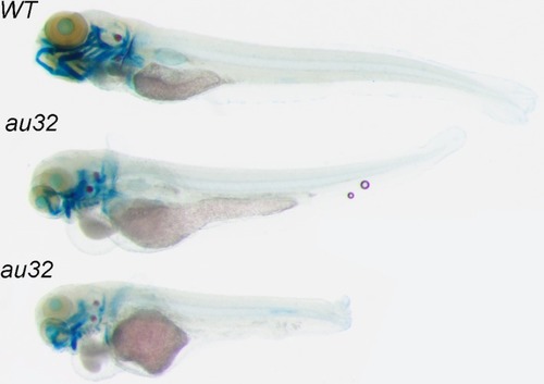
Whole body defects in PHENOTYPE:
|
Craniofacial defects in PHENOTYPE:
|

Unillustrated author statements |