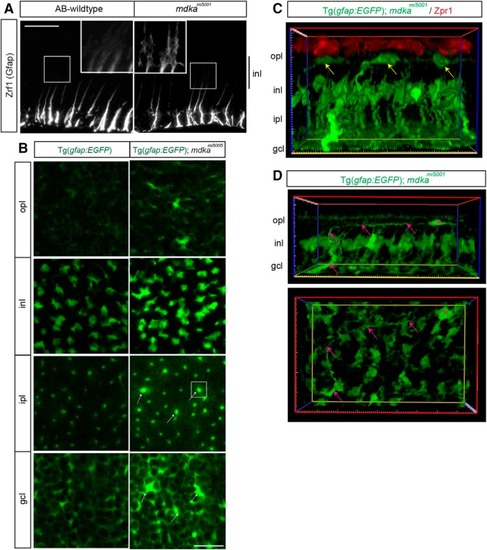
Following photoreceptor death, Müller glia in the mdkami5001 mutant undergo gliotic remodeling. A, Immunocytochemistry for Gfap in WT and mdkami5001 retinas at 28 dpl. In WT, the Gfap immunosignal is restricted to the inner third of radial processes. No obvious signal is detected at the inner nuclear layer. The mdkami5001 upregulates Gfap, and signals are seen at the cell body of Müller glia in the inner nuclear layer. B, Single optical planes from z-stack series of the Tg(gfap: EGFP) reporter flat-mount retinal preparation in the mdkami5001 background. In the ganglion cell and inner plexiform layers, some Müller glia show signs of hypertrophy, including increased levels of the EGFP transgene signal (arrows). C, Cross section view of 3D reconstructed image in the Tg(gfap:GFP); mdkami5001 (green) retina at 28 dpl, immunolabeled with Zpr1 (red) in a flat-mount preparation. Yellow arrows indicate displaced Müller glia somata in the outer plexiform layer. D, Cross section and flat-mounted views of the 3D reconstructed image. Displaced Müller glia (magenta asterisk) retain basal radial process (magenta arrows). opl, Outer plexiform layer; inl, inner nuclear layer; ipl, inner plexiform layer; gcl, ganglion cell layer. Scale bars: A, 30 μm; B, 20 μm.
|
