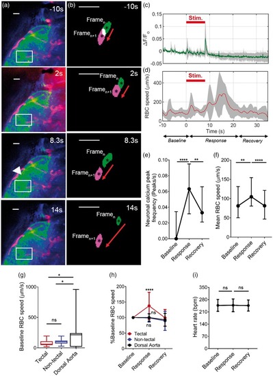
8 dpf zebrafish larvae display neurovascular coupling. (a) The left optic tectum of an 8 dpf Tg(nbt:GCaMP3; kdrl:mCherry;gata1:DsRED) embryo before (−10 s), during (2 s and 8.3 s) and after (14 s) visual stimulus by red light. Arrow indicates area of increased tectal calcium levels in response to the stimulus. (b) Segmented RBCs shown for two consecutive frames (Framen and Framen+1) for corresponding time point of the neuronal responses shown in (a). Individual RBCs are labelled in green to represent Framen and magenta to represent Framen+1. (c) Quantification of neuronal activation (ΔF/Fo) in optic tectum over time (n = 5 larvae). Visual stimulus was administered 0–8 s (indicated on graph). Timeseries was divided into baseline (−10 – 0 s), response (0–20 s), and recovery (20–30 s) periods. (d) Erythrocyte (RBC) speed in tectal vessels in the same animals as (c). (e) Quantification of frequency of peaks in (ΔF/Fo) as a measure of neuronal activation during baseline, response and recovery time periods (n = 40 larvae). (f) Mean RBC speed in the tectal vessels during baseline, response and recovery time periods in the same animals as (e). (g) Baseline RBC speed in tectal vessels, non-tectal (hindbrain and forebrain) vessels and dorsal aorta. (h) RBC speed expressed as percentage from the baseline for baseline, response and recovery for tectal vessels, non-tectal vessels and dorsal aorta (n = 17 larvae/group). (i) Beating heart rate (bpm) quantified for baseline, response and recovery time periods (n = 9 larvae/group). *p < 0.05, **p < 0.01, ****p < 0.0001. Scale bar represents 20 µm. Data in (e) and (f) are median and interquartile range (25% and 75% percentile). All other data are mean ± s.d.
|
