|
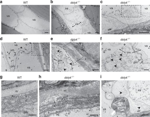
Ultrastructure of the notochord and notochord sheath in WT and <italic>dstyk</italic> mutant.a–f The transmission electron micrographs of the notochord in WT (a, d) and dstyk mutant (b, c, e and f) at 48 hpf. Note that the vacuoles were smaller in the dstyk mutant, and there were many scatteredly distributed small vesicles that were wrapped by single layer membrane in the cytoplasm of dstyk mutant. g, h The ultrastructure of the notochord sheath in the WT (g) and dstyk mutant (h). Note the straight, well-organized sheath layer in the WT and wavy, disordered organization of the medial layer in the dstyk mutant. i Ultrastructure of the notochord showed not well vacuolated epithelia-like cells (white arrow) aggregated in dstyk mutant. f, g and h were magnified image of the dotted boxes in c, d and e, respectively. Black arrowheads indicate many small vesicles in the mutant cytoplasm. All images are shown longitudinal sections. The scale bars represent (a–c, and i) 5 µm, (d–f) 1 µm and (g, h) 500 nm. va, vacuole; s, notochord sheath; sc, sheath cell; pm, plasma membrane; vm, vacuole membrane; i, inner laminin-rich layer, m, medial layer, and o, outer collagen-rich layers of the notochord sheath.
|