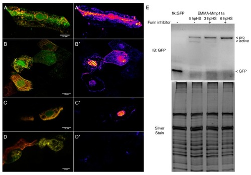
Mmp11a is activated by furin except in the nucleus. (A) Myocytes expressing EMMA–Mmp11a remove the hemagglutinin (HA)-tagged propeptide (red channel), leaving only the carboxyl terminal GFP tag (green channel) as the construct traverses the secretory pathway, and also in the nucleus. This is more clearly visible in the false color heat map showing the strength of the GFP signal relative to the HA signal (A′). (B) Epithelial cells expressing EMMA–Mmp11a also remove the propeptide as the construct is secreted and released extracellularly, as well as in the nucleus. (B′) Heat maps of the relative abundance of GFP vs. HA clearly illustrate the activation of the EMMA–Mmp11a construct as it is secreted, as well as within the nuclei. (C) Myocytes expressing EMMA–Mmp11a in the presence of furin inhibitor only remove the propeptide in the nuclei. (C′) Heat maps of myocytes expressing the construct in the presence of the furin inhibitor show relatively unimpaired proteolytic removal of the propeptide in the nuclei, but dramatically reduced activation in the secretory pathway and extracellularly. (D) Epithelial cells expressing the construct in the presence of furin inhibitor also show dramatically reduced activation. (D′) Heat maps of epithelial cells in the presence of furin inhibitor, showing negligable activation of the contstruct. (E) Immunoblots of embryo homogenates expressing either GFP alone (flk:GFP), or EMMA–Mmp11a, in the presence or absence of furin inhibitor, probed with anti-GFP. Activation of the construct is clearly detectable in embryos 6 h post heat shock (hpHS) in the absence of inhibitor. In the presence of inhibitor, some activation is detectable 3 hpHS, but by 6 hpHS activation, appears completely abolished. Silver staining of a replicate gel shows comparable protein loads. Scale bars are 10 µm.
|
