FIGURE

Figure 1

ID
ZDB-FIG-200125-55
Publication
Matchett et al., 2019 - Paralogues of Mmp11 and Timp4 Interact during the Development of the Myotendinous Junction in the Zebrafish Embryo
Other Figures
All Figure Page
Back to All Figure Page
Figure 1

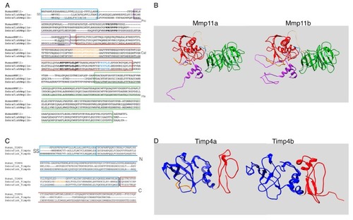
Sequences and predicted structures of zebrafish metalloproteinase 11 (Mmp11) and tissue inhibitors of metalloproteinase-4 (Timp4) paralogues exhibit both conserved and divergent features. (A) Inferred amino acid sequences of zebrafish Mmp11a and Mmp11b paralogues aligned with human MMP11 for reference. Secretory signals are boxed in light blue, propeptide in purple (with cysteine switch motif highlighted in bold and furin recognition sequence underlined), catalytic domain in red (with S-loop in orange, zinc binding motif in bold italic, and specificity loop in light blue), and the carboxyl hemopexin domain is boxed in green. (B) Structural homology models of zebrafish Mmp11a and Mmp11b rendered as ribbon diagrams and colored as in the sequence alignment. (C) Inferred amino acid sequences of zebrafish Timp4a and Timp4b paralogues aligned with human TIMP4 for reference. Secretory signals are boxed in light blue, the N-terminal domain in blue (with the seven-residue charged insertion of Timp4a highlighted in orange), and the C-terminal domain in red. (D) Structural homology models of zebrafish Timp4a and Timp4b rendered as ribbon diagrams and colored as in the sequence alignment.

Expression Data

Expression Detail
Antibody Labeling
Phenotype Data

Phenotype Detail
Acknowledgments
This image is the copyrighted work of the attributed author or publisher, and ZFIN has permission only to display this image to its users. Additional permissions should be obtained from the applicable author or publisher of the image. Full text @ J Dev Biol