|
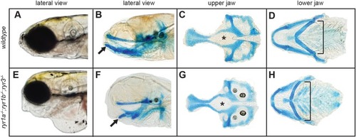
Paralyzed ryr mutants have abnormal craniofacial morphology. Craniofacial features of 6 dpf larvae were visualized following staining with Alcian Blue, which marks cartilage. (A-D) Jaw structures of wild-type larvae: lateral view of intact larva (A), lateral view of stained intact larva (B), isolated upper jaw (C) and isolated lower jaw (D). (E-H) Jaw structures of paralyzed ryr1a;ryr1b;ryr3 larvae: lateral view of intact larva (E), lateral view of stained intact larva (F), isolated upper jaw (G) and isolated lower jaw (H). Jaw architecture of mutant larvae was dramatically shortened in the A-P dimension and wider than normal. Highlighted features include the shape of hypophyseal fenestre (asterisks), the angle between the midline and ceratohyal cartilages of the lower jaw (brackets) and orientation of Meckel's cartilage (arrows).
|