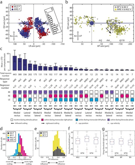
Monocular/binocular synopsis. a Transversal projection of monocular coding neurons within rh5/6 (ABN). D, dorsal; L, left; M, Mauthner cells; MLE, monocular left eye; MRE, monocular right eye; P, position; R, right; V, ventral. Black arrows indicate position of a faint gap between the ventral and dorsal clusters of putative internuclear neurons. Inset shows the numbers of neurons plotted in this figure for the left hemisphere along the D-V axis rotated by 20°. b Monocular and binocular velocity encoding neurons. A, anterior; BA, binocular always; BP, binocular preferred; P, posterior; rh 5-8, rhombomere 5-8; V, velocity. Black arrow indicating the direction of the velocity shift. c Sum of the total number of neurons found for each response type sorted pairwise according to the affected muscle(s). The bar plot shows the mean and standard deviation for eight composite brains. BA, binocular always; BP, binocular preferred; MLE, monocular left eye; MLEX, monocular left eye exclusive; MRE, monocular right eye; MREX, monocular right eye exclusive. d Monocular coding differences for all four main response types for position coding neurons. Index running from − 1 (exclusively coding for left eye) to + 1 (right eye). e PV influence for BA P and BP P neurons. Index running from − 1 (exclusive velocity influence) to + 1 (exclusive position influence). f, g Left and right eye firing thresholds acquired during the firing threshold analysis pooled in ON direction
|
