
FIGURE
Fig. 4
- ID
- ZDB-FIG-191230-1489
- Publication
- Hamada et al., 2019 - Pattern of fin rays along the antero-posterior axis based on their connection to distal radials
- Other Figures
- All Figure Page
- Back to All Figure Page
Fig. 4
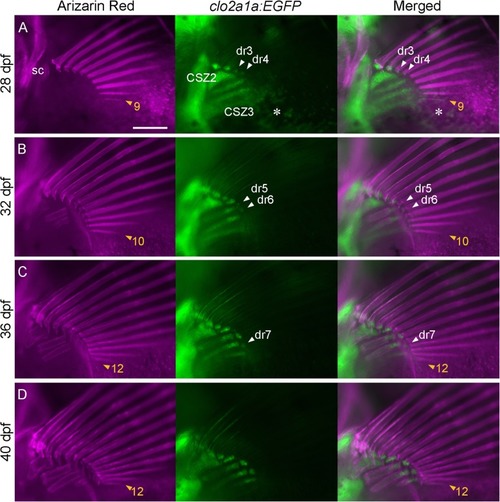
Process of morphogenesis of the fin rays and radials. Calcified bones (Alizarin Red) and chondrogenic cells ( |
Expression Data
Expression Detail
Antibody Labeling
Phenotype Data
Phenotype Detail
Acknowledgments
This image is the copyrighted work of the attributed author or publisher, and
ZFIN has permission only to display this image to its users.
Additional permissions should be obtained from the applicable author or publisher of the image.
Full text @ Zoological Lett