IMAGE

Fig. 4

ID
ZDB-IMAGE-191230-1491
Source
Figures for Hamada et al., 2019
Image
Figure Caption

Fig. 4

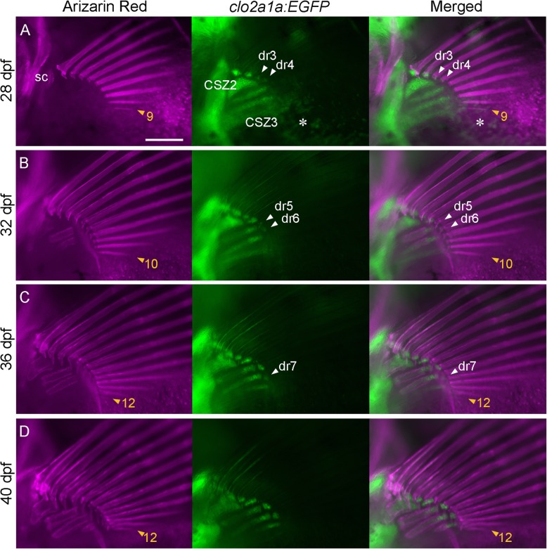
Process of morphogenesis of the fin rays and radials. Calcified bones (Alizarin Red) and chondrogenic cells (col2a:EGFP) of the left pectoral fin in individual tracing specimens were observed at 28 dpf (a), 32 dpf (b), 36 dpf (c), and 40 dpf (d). The observed fish were crossbred. Orange arrowheads with numbers indicate the most posterior fin ray. White arrowheads with dr3–7 indicate newly appearing distal radials. The scale bar in a indicates 200 μm. CSZ, cartilage subdivision zone. sc, anlagen of the scapula. The asterisk in a indicates the reflected signal of the iridophore on the swim bladder

Acknowledgments
This image is the copyrighted work of the attributed author or publisher, and ZFIN has permission only to display this image to its users. Additional permissions should be obtained from the applicable author or publisher of the image. Full text @ Zoological Lett