
Fig. 4
- ID
- ZDB-FIG-191028-4
- Publication
- Dallinga et al., 2018 - IGF2 and IGF1R identified as novel tip cell genes in primary microvascular endothelial cell monolayers
- Other Figures
- All Figure Page
- Back to All Figure Page
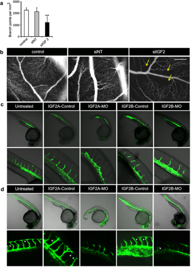
IGF2 is essential for sprouting in the CAM model and zebrafish embryos. a Quantification of number of branching points/mm2 comparing untreated membranes and membranes treated with siNT or with siIGF2 in the CAM model. ***p < 0.001 as compared to control as well as siNT treatment. b Representative images of the vascular network in CAMs of chicks that were treated with siNT or siIGF2 and untreated control. Arrows indicate non-vascularized areas in the vascular network. Scale bar represents 500 µm. c, dRepresentative images of Tg(fli1a-eGFP) zebrafish embryos at 24 h (c) and 30 h (d) after injection of either a Morpholino targeting Igf2a (IGF2A-MO) or Igf2b (IGF2B-MO) or a 6-bp mismatch control Morpholino for each gene (IGF2A-CON and IGF2B-CON, respectively). Untreated zebrafish embryos are shown as a control. Arrowheads indicate filopodia |
| Gene: | |
|---|---|
| Fish: | |
| Knockdown Reagents: | |
| Anatomical Term: | |
| Stage Range: | Prim-5 to Prim-15 |
| Fish: | |
|---|---|
| Knockdown Reagents: | |
| Observed In: | |
| Stage Range: | Prim-5 to Prim-15 |