
Fig. 6
- ID
- ZDB-FIG-190913-72
- Publication
- Toms et al., 2019 - Phagosomal and mitochondrial alterations in RPE may contribute to KCNJ13 retinopathy
- Other Figures
- All Figure Page
- Back to All Figure Page
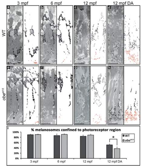
Melanosome localization in the obetd15 retinal pigment epithelium (RPE) in normal and dark-adapted conditions. TEM images of wild-type (WT) (a–d) and obetd15 (e–h) RPE at 3 months post fertilization (mpf), 6 mpf and 12 mpf with neighbouring panels showing digitally extracted melanosomes with the basal RPE-localized melanosomes false-colored in red. Using these extracted melanosomes, the proportion of melanosomes localized to the photoreceptor region was calculated, shown in bar chart (i). There was little difference in the proportion of melanosomes localised to the apical photoreceptor region of the RPE in zebrafish exposed to a normal daily light cycle. Whereas, dark-adaptation (DA) of 12 mpf obetd15 zebrafish (h) caused significantly more melanosomes to localize to the basal region of the RPE compared to WT zebrafish eyes (d). Results are the mean (from three regions from three eyes) ± SEM. *p < 0.05. Scale bars = 10 µm. |
| Fish: | |
|---|---|
| Condition: | |
| Observed In: | |
| Stage: | Adult |