
Fig. 3
- ID
- ZDB-FIG-190913-59
- Publication
- Frikstad et al., 2019 - A CEP104-CSPP1 Complex Is Required for Formation of Primary Cilia Competent in Hedgehog Signaling
- Other Figures
- All Figure Page
- Back to All Figure Page
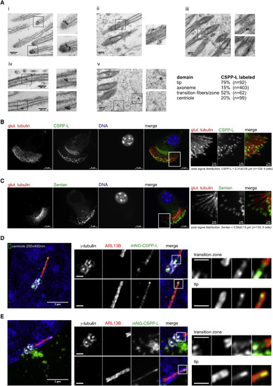
Ciliary Localization of CSPP-L at Motile and Primary Cilia (A) CSPP-L detection by post-embedding IEM of mouse tracheal epithelial cells. Panels depict close ups of (i) cilia tips, (ii and iii) basal bodies, (iv) cilia axonemes, and (v) apically localized electron-dense particles. (B and C) IFM of mouse tracheal epithelial cells showing axonemal MTs (glutamylated tubulin, red) and CSPP-L (B, green) or Sentan (C, green). Right panels show magnifications of indicated regions. (D and E) 3D-SIM IFM of hTERT-RPE1 cells expressing monomeric NeonGreen (mNG)-CSPP-L and co-stained for centrosomal marker γ-tubulin (white) and cilia membrane marker ARL13B (red). Scale bars in magnified areas, 500 nm. mNG-CSPP-L decorates axonemal MTs throughout the transition zone and concentrates at the tip (D and E). Centriolar satellite localization is frequently found and exemplified in (E) and Figure S4C. |