
Fig. S1
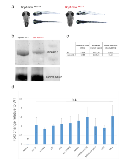
mok m632−/− embryo morphology at 6dpf, Dynactin1 protein quantification at 2dpf and qRT-PCR expression in mok m632−/− larvae. a) Wild-type sibling and homozygous mutant embryo morphology at 6dpf; close-up showing a dorsal view of the head to emphasize previously described eye phenotype. b) Western blot of maternally-contributed Dynactin1 in 2dpf mok m632−/− embryo (detected with anti-DCTN1 antibody from Origene, TA346929), c) quantified against gamma-tubulin at 32% wild-type level. d) Quantification of 3 biological replicates of qRT-PCR levels from 6dpf mok m632−/− larvae mRNA relative to the average wild-type levels obtained for 6dpf m632+/+ larvae mRNA (presented as fold change) shows no compensation by dynctin1b or kif14, no change in the expression of other subunits of the dynactin complex (p22/24, p25, p50, actr1), no change in other known regulators of the dynein motor complex (ndel1b, pafah1b1a/1b1b), and no changes indicative of trophic compensation (bdnf). (TIF 45716 kb) |