
Fig. 5
- ID
- ZDB-FIG-190816-10
- Publication
- Kuroda et al., 2018 - Roles of basal keratinocytes in actinotrichia formation
- Other Figures
- All Figure Page
- Back to All Figure Page
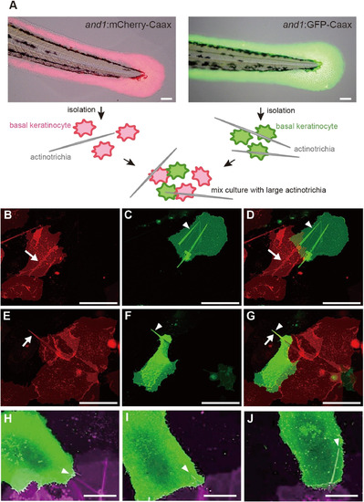
In vitro holding of the long mature actinotrichia by the basal keratinocytes from two reporter lines. (A) Schematic diagram of the mix-culture experiment using two types of reporter zebrafish. Scale bar is 50 μm. (B–J) The cultured basal keratinocytes in vitro from the larvae of and1:mCherry-CaaX and and1:GFP-CaaX. (B) White arrow indicates the actinotrichium in the basal keratinocyte expressing mCherry-Caax. (C) White arrowhead indicates the actinotrichium in the basal keratinocyte expressing GFP-CaaX. (E) White arrow indicates the actinotrichium protruded from a basal keratinocyte were enveloped by a mCherry-CaaX-expressing basal keratinocyte. (F) White arrowhead indicates GFP-CaaX-expressing basal keratinocyte. (D and G) Merged image of (B and C) and (E and F), respectively. Two adjacent basal keratinocytes share the same actinotrichia. Scale bar in (B − G) is 50 μm. (H–J) Time-lapse images of the plasma membrane enveloping the actinotrichium. White arrowheads indicate the actinotrichium enveloped by two adjacent basal keratinocytes. Scale bar in (H–J) is 20 μm. |
Reprinted from Mechanisms of Development, 153, Kuroda, J., Iwane, A.H., Kondo, S., Roles of basal keratinocytes in actinotrichia formation, 54-63, Copyright (2018) with permission from Elsevier. Full text @ Mech. Dev.