
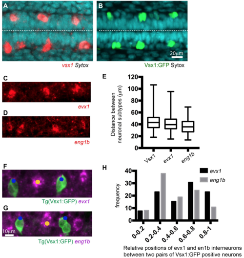
Fig. S3
Neuronal spacing pattern in the zebrafish spinal cord (related to Figure 4). A) and B) Dorsal views of Vsx1 expressing cells revealed by in situ hybridization (A) and Vsx1:GFP transgene expression (B) at 20 hfp. Dashed line shows the apical surface. C) and D) Lateral views of evx1 (C) and eng1b (D) expressing spinal neurons revealed by in situ hybridization at 20hpf. E) Box-and-whisker plot showing the distance between neurons of the same subtype in the most caudal spinal region at 20hpf (mean ± SD; Vsx1:GFP: 44.4 ± 14.1 µm, n=18; evx1: 41 ± 15.4 µm, n=7; eng1b: 35.7 ± 13.3 µm, n=5 embryos). The line inside the box represents the median and whiskers represent minimum and maximum values. Data analysed using Kruskal-Wallis with Dunn’s multiple comparison test (non-significant). F and G) Dorsolateral views showing the relative positions of Vsx1:GFP and evx1 (F) or eng1b (G) expressing cells revealed by in situ hybridisation. H) Frequency distribution chart shows the relative positions of evx1 and en1b expressing interneurons between two pairs of Vsx1:GFP expressing neurons. The distance between the two pairs of Vsx1:GFP neurons has been normalised from 0 (anterior) to 1 (posterior). All evx1 and eng1b expressing neurons that shared the same position with a Vsx1 neuron have been included in position (1). |
Reprinted from Developmental Cell, 49, Hadjivasiliou, Z., Moore, R.E., McIntosh, R., Galea, G.L., Clarke, J.D.W., Alexandre, P., Basal Protrusions Mediate Spatiotemporal Patterns of Spinal Neuron Differentiation, 907-919.e10, Copyright (2019) with permission from Elsevier. Full text @ Dev. Cell