FIGURE

Figure 6

ID
ZDB-FIG-190723-450
Publication
Hadjivasiliou et al., 2019 - Basal Protrusions Mediate Spatiotemporal Patterns of Spinal Neuron Differentiation
Other Figures
All Figure Page
Back to All Figure Page
Figure 6

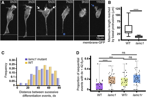
Laminin Depletion Reduces Basal Protrusion Length and Spacing between Successively Differentiating Neurons

(A) Time-lapse sequence showing a differentiating neuron in a Laminin-depleted spinal cord (see Video S6). It has only short basal protrusions (white arrows in time point 5h00). The short basal protrusions are retracted before detachment from apical surface (blue asterisk at 7h00) and axon initiation (blue arrow at 11h00). Cell is labeled with membrane-GFP. View is dorsal.

(B) Box-and-whisker plot showing the maximum length reached by basal protrusions in wild-type (mean ± SD, 42.6 ± 20.2 μm, n = 24 cells) and lamc1-mutant embryos (mean ± SD, 12.3 ± 4.7 μm, n = 39 cells). The line inside the box represents the median and whiskers represent minimum and maximum values. Data analyzed using unpaired one-tailed Mann-Whitney test (p-value < 0.0001).

(C) Histogram showing the distribution of the distance between successive Vsx1:GFP differentiation events in wild-type (orange) and lamc1-mutant embryos (purple) (mean ± SD, 54.00 ± 1.52 μm in wild-type and 45.3 ± 0.99 μm in lamc1, one-tailed t test p-value = 3.16 10−6).

(D) Graph showing the proportion of successive Vsx1:GFP differentiation events that occur within a 42.6-μm interval (the average size of wild-type basal protrusions) in wild-type embryos, lamc1 embryos, randomized wild-type distributions and randomized lamc1 distributions. The Kolmogorov-Smirnov test was used to compare wild-type and lamc1 distributions (p-value = 0.000066); wild-type and randomized wild-type distributions (p = 0.000224); and, lamc1 and randomized lamc1 distributions (p-value = 0.213).

Expression Data

Expression Detail
Antibody Labeling
Phenotype Data

Phenotype Detail
Acknowledgments
This image is the copyrighted work of the attributed author or publisher, and ZFIN has permission only to display this image to its users. Additional permissions should be obtained from the applicable author or publisher of the image.

Reprinted from Developmental Cell, 49, Hadjivasiliou, Z., Moore, R.E., McIntosh, R., Galea, G.L., Clarke, J.D.W., Alexandre, P., Basal Protrusions Mediate Spatiotemporal Patterns of Spinal Neuron Differentiation, 907-919.e10, Copyright (2019) with permission from Elsevier. Full text @ Dev. Cell