
Fig. 2
- ID
- ZDB-FIG-190812-13
- Publication
- Kuwata et al., 2019 - Local heat-shock mediated multi-color labeling visualizing behaviors of enteric neural crest cells associated with division and neurogenesis in zebrafish gut
- Other Figures
- All Figure Page
- Back to All Figure Page
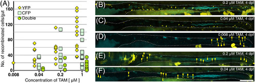
The number and distribution of ENCCs recombined in the gut at different concentrations of TAM. We treated heat‐shocked Tg(hsp:cre) x Tg (ubi:ZB) embryos with different concentrations of TAM, and counted the YFP+, CFP+, and YFP/CFP‐double+ cells in the left side of the gut at ca. 110 hpf. A, The number of recombinant ENCCs in the gut detected by YFP or CFP was dependent on the TAM concentration. More YFP+ cells were detected than CFP+ cells, probably because of the weaker signal of CFP. Numbers of RFP+ embryos examined for each concentration are 11 for 0.008 μM, 17 for 0.04 μM, 19 for 0.2 μM, and 18 for 1 μM. B‐F, Representative examples from each TAM (0.008‐0.2 μM) treatment. Samples E and F show the smallest number of YFP+ cells at each concentration of TAM. Some groups of cells were localized, whereas others were distributed along the anterior–posterior axis of the gut. White dotted lines outline the guts. Yellow triangles: YFP+ cells. Scale bar = 100 μm in F |