Kuwata et al., 2019 -
Local heat-shock mediated multi-color labeling visualizing behaviors of enteric neural crest cells associated with division and neurogenesis in zebrafish gut
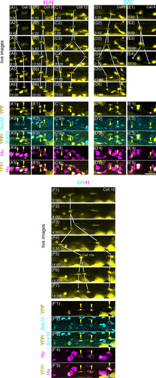
Process of cell division of each ENCC and the state of differentiation of its daughter cells revealed by time‐lapse imaging and post‐hoc immunostaining. A1‐F7, Serial images of time‐lapse movies of six dividing cells. The cell ID number, shown at the top panel of each column, corresponds to those in Figure 4. Cell nos. 3, 9, and 12 produced only Hu+/Sox10− daughter cells. Cell nos. 2 and 6 produced only Hu−/Sox10+ cells. Cell no. 10 is an example in which the second division was observed. The time after the starting point of each time‐lapse is shown at the bottom‐left corner of the panel. Each cell and its daughter cells at different time points are connected with solid lines. The daughter cells examined are marked with white dots. The left daughter cell in (D4) is outlined with dotted line. A′1‐F′5, Identification of the state of differentiation of daughter cells by immunostaining. Yellow triangles: daughter cells. Markers are indicated on the left of each row. The HuC/D signal in YFP+ daughter cells in A4′ is weak, although distinct from the signal in Hu−/Sox10− cell indicated with an arrow. The Sox10+ cell marked with a white asterisk in F2′ and F3′ was not Hu+, because it was in different confocal slices from Hu + cells in F′4 (data not shown). Sox10+ cell with a black asterisk in F′2 and F′3 was in the epidermal level. The positions of cells with no staining are encircled with dashed lines. Lateral view of the gut. Anterior, to the left. Dorsal, to the top. Scale bar = 25 μm
Expression Data
Expression Detail
Antibody Labeling
Phenotype Data
Phenotype Detail
Acknowledgments
This image is the copyrighted work of the attributed author or publisher, and
ZFIN has permission only to display this image to its users.
Additional permissions should be obtained from the applicable author or publisher of the image.
Full text @ Dev. Dyn.
Your Input Welcome
Thank you for submitting comments. Your input has been emailed to ZFIN curators who may contact you if
additional information is required.
Oops. Something went wrong. Please try again later.