
FIGURE
Fig. 2
- ID
- ZDB-FIG-190809-25
- Publication
- Crilly et al., 2019 - Using Zebrafish Larvae to Study the Pathological Consequences of Hemorrhagic Stroke
- Other Figures
- All Figure Page
- Back to All Figure Page
Fig. 2
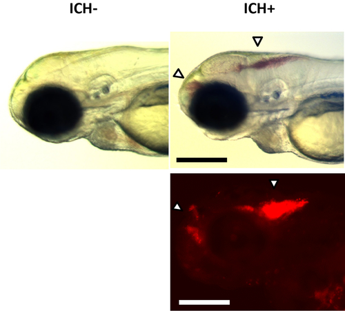
ICH+ brain hemorrhage phenotypes. Examples of larval ICH phenotypes maintained on a transgenic gata1:DsRed reporter nacre background observed with a brightfield stereomicroscope (top panels) and fluorescence (bottom panel) at ~48 h post-fertilization. No hemorrhages were observed in ICH-larvae (left panels). A distinct accumulation of red blood cells in the forebrain and hindbrain (arrows) were observed in ICH+ larvae (right panels). Scale bars represent 250 µm. Figure has been reproduced from Crilly et al.20 with permission under a Creative Commons license.
|
Expression Data
Expression Detail
Antibody Labeling
Phenotype Data
Phenotype Detail
Acknowledgments
This image is the copyrighted work of the attributed author or publisher, and
ZFIN has permission only to display this image to its users.
Additional permissions should be obtained from the applicable author or publisher of the image.
Full text @ J. Vis. Exp.