
Fig. 3
- ID
- ZDB-FIG-190806-36
- Publication
- Baek et al., 2019 - The Alternative Splicing Regulator Nova2 Constrains Vascular Erk Signaling to Limit Specification of the Lymphatic Lineage
- Other Figures
- All Figure Page
- Back to All Figure Page
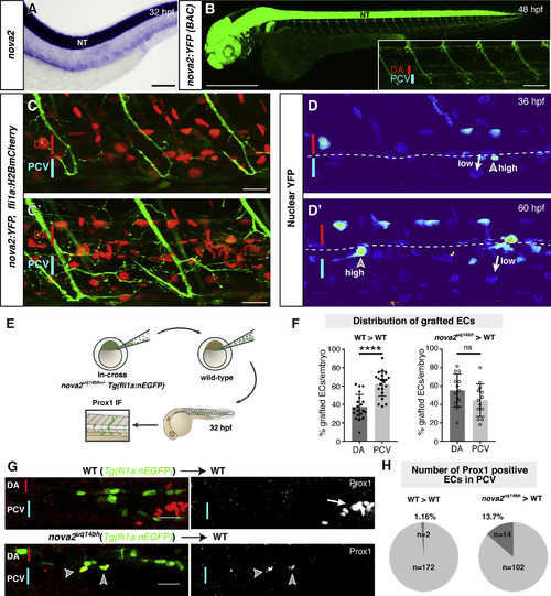
Nova2 Acts Cell Autonomously in Endothelial Cells (A) Expression of nova2 in the zebrafish trunk at 32 hpf by ISH. NT, neural tube. (B) Overview of Tg(nova2:YFP)uq40bh BAC transgenic line at 48 hpf. Inset = region of trunk vasculature displaying positive neurons, DA and PCV. (C) Tg(nova2:YFPuq40bh; fli1a:H2BmCherryuq37bh) double transgenic line at 36 hpf (C) and 60 hpf (Cʹ). (D) Nuclear Tg(nova2:YFP)uq40bh signal. H2BmCherry signal was used as a mask to limit analysis to the nucleus. Representative of n = 10 embryos analyzed at 36 hpf (D) and 60 hpf (Dʹ). (E) Schematic illustration representing the cell transplantation assay followed by Prox1 IF analysis. (F) Contribution of transplanted ECs to arterial and venous endothelial lineages. (G) Tg(fli1a:nEGFP)y7 donor cells derived from a wild-type (WT, top) or nova2uq14bh mutant (bottom) grafted into a WT acceptor embryo analyzed by Prox1 IF. Arrowheads indicate Prox1 and GFP double-positive mutant nuclei in the PCV. Arrow indicates Prox1-positive corpuscle of Stannius (CS). (H) Quantification of the percentage of Prox1 and GFP double-positive cells located in the dorsal side of PCV at 32 hpf (number of cells shown from sibling grafts (n = 21 grafts) and mutant grafts (n = 13 grafts) into wild-type hosts). Scale bars: 100 μm in (A), 500 μm in (B), 50 μm in inset figure, 20 μm in (C) and (Cʹ), 25 μm in (G). Error bars: SD, Mann-Whitney-test, ∗∗∗∗p < 0.0001, ns: not significant. See also Figure S3. |
| Genes: | |
|---|---|
| Fish: | |
| Anatomical Terms: | |
| Stage Range: | Prim-15 to Pec-fin |
Reprinted from Developmental Cell, 49, Baek, S., Oh, T.G., Secker, G., Sutton, D.L., Okuda, K.S., Paterson, S., Bower, N.I., Toubia, J., Koltowska, K., Capon, S.J., Baillie, G.J., Simons, C., Muscat, G.E.O., Lagendijk, A.K., Smith, K.A., Harvey, N.L., Hogan, B.M., The Alternative Splicing Regulator Nova2 Constrains Vascular Erk Signaling to Limit Specification of the Lymphatic Lineage, 279-292.e5, Copyright (2019) with permission from Elsevier. Full text @ Dev. Cell