
Fig. 1
- ID
- ZDB-FIG-190806-34
- Publication
- Baek et al., 2019 - The Alternative Splicing Regulator Nova2 Constrains Vascular Erk Signaling to Limit Specification of the Lymphatic Lineage
- Other Figures
- All Figure Page
- Back to All Figure Page
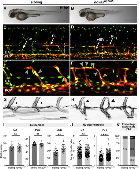
nova2uq14bh Mutants Exhibit Increased Lymphatic Sprouting (A and B) Overall morphology of a sibling (A) and nova2uq14bh mutant (B) at 54 hpf. (C–F) Trunk vasculature labeled with Tg(fli1a:nEGFP)y7;Tg(5.2lyve1b:DsRed)nz101 in a sibling (C) and (E) and a nova2uq14bh mutant (D) and (F) at 54 hpf (venous and LECs in red). Arrowheads indicate LEC nuclei. Venous intersegmental vessel (vISV) and PL. (G and H) Tg(fli1ep:lifeact-EGFP) labeling endothelial F-actin in PLs of a sibling (G) and a nova2uq14bh mutant (H) at 54 hpf. Arrowheads show exploratory PLs. (I) EC numbers within different lineages: dorsal aorta (DA, arterial), posterior cardinal vein (PCV, venous), and lymphatic ECs (LECs) at 54 hpf. Quantification across 5 body segments (siblings n = 17, mutants n = 17). (J) Quantification of nuclear ellipticity of ECs in the DA and PCV at 54 hpf. Quantification across 5 body segments (siblings n = 11, mutants n = 11). (K) Quantification of hyperactive PLs (defined as displaying cellular extensions that probe dorsal to the horizontal myoseptum [HM]) in siblings and nova2uq14bh mutants across 3 body segments (siblings n = 10, mutants n = 10). Light gray indicates wild-type morphology, dark gray indicates exploratory PLs in 1/2 body segments scored, and black indicates exploratory PLs in 2/2 body segments scored (p = 0.0174). Error bars represent SD, t test (I), or Mann-Whitney-test (J) and (K), ∗∗p < 0.01, ∗∗∗∗p < 0.0001, and ns; not significant; Scale bar represents 1 mm in (A) and (B), 25 μm in (C)–(F), and 20 μm in (G) and (H). See also Figures S1 and S2.
|
| Genes: | |
|---|---|
| Fish: | |
| Anatomical Terms: | |
| Stage: | Long-pec |
| Fish: | |
|---|---|
| Observed In: | |
| Stage Range: | Long-pec to Day 5 |
Reprinted from Developmental Cell, 49, Baek, S., Oh, T.G., Secker, G., Sutton, D.L., Okuda, K.S., Paterson, S., Bower, N.I., Toubia, J., Koltowska, K., Capon, S.J., Baillie, G.J., Simons, C., Muscat, G.E.O., Lagendijk, A.K., Smith, K.A., Harvey, N.L., Hogan, B.M., The Alternative Splicing Regulator Nova2 Constrains Vascular Erk Signaling to Limit Specification of the Lymphatic Lineage, 279-292.e5, Copyright (2019) with permission from Elsevier. Full text @ Dev. Cell