
FIGURE
Fig. S3
- ID
- ZDB-FIG-190805-13
- Publication
- Herzog et al., 2019 - Rapid clearance of cellular debris by microglia limits secondary neuronal cell death after brain injury in vivo
- Other Figures
- All Figure Page
- Back to All Figure Page
Fig. S3
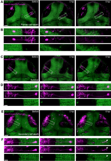
In vivo imaging of individual tectal neurons shows that both primary and secondary cell death occur after brain injury. (A,C,E) Confocal images of theoptic tectum of H2A:GFP transgenic animals, where tectal neurons labelled through injection of elavl3:memTdTomato plasmid DNA die through primary cell death (A), survive (C), or die through secondary cell death (E) after mechanical injury. Scale bars, 50 μm. (B,D,F) Close-up of neurons indicated in (A), (C) and (E). White arrow indicates pyknotic nucleus. Scale bars, 10 μm. |
Expression Data
Expression Detail
Antibody Labeling
Phenotype Data
Phenotype Detail
Acknowledgments
This image is the copyrighted work of the attributed author or publisher, and
ZFIN has permission only to display this image to its users.
Additional permissions should be obtained from the applicable author or publisher of the image.
Full text @ Development