
FIGURE
Fig. S2
- ID
- ZDB-FIG-190805-12
- Publication
- Herzog et al., 2019 - Rapid clearance of cellular debris by microglia limits secondary neuronal cell death after brain injury in vivo
- Other Figures
- All Figure Page
- Back to All Figure Page
Fig. S2
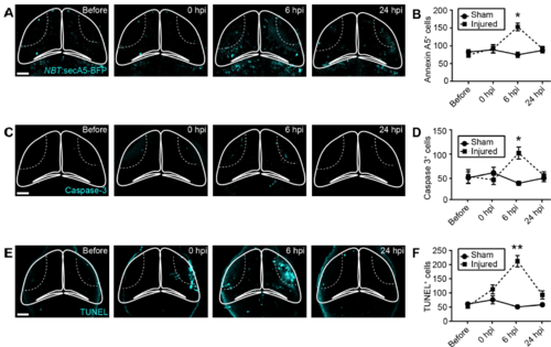
Cell death after brain injury can be detected through Annexin A5 live imaging, caspase 3 immunohistochemistry, and TUNEL staining. (A,C,E) Confocal images of the tectum of a NBT:secA5-BFP animal (A), and animals after cleaved caspase 3 immunohistochemistry (B) or TUNEL staining (C), at different time points after injury. Scale bars, 50 μm. (B,D,F) Quantification of Annexin A5+ cells (B), cleaved caspase 3+ cells (D) or TUNEL+ cells (F). n ≥ 6 animals per experimental group. *, p < 0.05 and **, p<0.01 in two-way ANOVA |
Expression Data
Expression Detail
Antibody Labeling
Phenotype Data
Phenotype Detail
Acknowledgments
This image is the copyrighted work of the attributed author or publisher, and
ZFIN has permission only to display this image to its users.
Additional permissions should be obtained from the applicable author or publisher of the image.
Full text @ Development