|
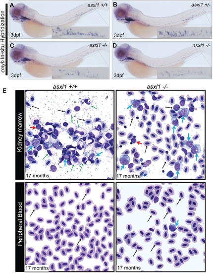
Complete loss of asxl1 leads to AML in zebrafish. WISH for cmyb was performed at 3 dpf in asxl1+/+ (A), asxl1+/− (B) and asxl1−/− (C) zebrafish embryos obtained from a cross of two asxl1+/− fish. (D) WISH for cmyb was performed at 3 dpf in asxl1−/− zebrafish embryos obtained from a cross of two asxl1−/− fish. Insets show a higher magnification of the CHT in each panel. (E) MGG staining was performed on kidney marrow smears and peripheral blood smears of asxl1+/+ and asxl1−/− fish at 17 months of age. Normal maturation and morphology are shown across a spread of blood cells in the kidney marrow of the asxl1+/+ zebrafish. In the asxl1−/− fish, the kidney marrow is replaced with immature myeloid blast cells in a pattern resembling AML. Circulating immature myeloid blast cells were observed in the peripheral blood of the asxl1−/− zebrafish, but not asxl1+/+ fish. Erythrocytes, black arrow; myeloid cells, green arrow; blast cells, blue arrow; lymphocytes, red arrow.
|