IMAGE

Fig. 4.

ID
ZDB-IMAGE-190723-920
Genes
Source
Figures for Gjini et al., 2019
Image
Figure Caption

Fig. 4.

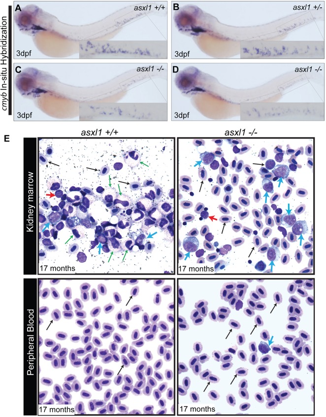
Complete loss of asxl1 leads to AML in zebrafish. WISH for cmyb was performed at 3 dpf in asxl1+/+ (A), asxl1+/ (B) and asxl1−/− (C) zebrafish embryos obtained from a cross of two asxl1+/ fish. (D) WISH for cmyb was performed at 3 dpf in asxl1−/− zebrafish embryos obtained from a cross of two asxl1−/− fish. Insets show a higher magnification of the CHT in each panel. (E) MGG staining was performed on kidney marrow smears and peripheral blood smears of asxl1+/+ and asxl1−/− fish at 17 months of age. Normal maturation and morphology are shown across a spread of blood cells in the kidney marrow of the asxl1+/+ zebrafish. In the asxl1−/− fish, the kidney marrow is replaced with immature myeloid blast cells in a pattern resembling AML. Circulating immature myeloid blast cells were observed in the peripheral blood of the asxl1−/− zebrafish, but not asxl1+/+ fish. Erythrocytes, black arrow; myeloid cells, green arrow; blast cells, blue arrow; lymphocytes, red arrow.

Figure Data
Acknowledgments
This image is the copyrighted work of the attributed author or publisher, and ZFIN has permission only to display this image to its users. Additional permissions should be obtained from the applicable author or publisher of the image. Full text @ Dis. Model. Mech.