
FIGURE
Figure 1 - figure supplement 2
- ID
- ZDB-FIG-190723-782
- Publication
- Hardy et al., 2019 - Detailed analysis of chick optic fissure closure reveals Netrin-1 as an essential mediator of epithelial fusion
- Other Figures
- All Figure Page
- Back to All Figure Page
Figure 1 - figure supplement 2
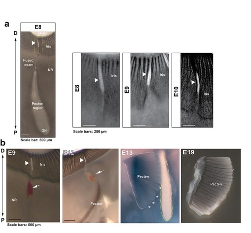
Anatomical features of iris and pecten in relation to OFC in the chick eye. ( |
Expression Data
Expression Detail
Antibody Labeling
Phenotype Data
Phenotype Detail
Acknowledgments
This image is the copyrighted work of the attributed author or publisher, and
ZFIN has permission only to display this image to its users.
Additional permissions should be obtained from the applicable author or publisher of the image.
Full text @ Elife