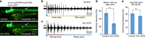
V1 ablation reduced cycle frequency in swimming. a Fluorescent images (green channel) of Tg[en1b:GFP] and Tg[en1b:loxP-RFP-loxP-DTA] fish with (bottom panel) or without (top panel) Tg[hoxa4a/9a:Cre]. Green fluorescent protein (GFP) expression in the spinal V1 neurons (arrow) is absent in the presence of Cre (En1-DTA), with GFP expression in the brain (triangle) and slow muscle cells in the middle region of the body (arrowhead) being intact. Scale bar, 250 μm. b, c Ventral root recordings of fictive swimming elicited by electrical stimulations (ESs) in a control (b) and an En1-DTA fish (c). d Swimming frequency of control and En1-DTA fish during the initial phase of ES swim (swimming elicited by electrical stimulation). Control: 54.8 ± 5.7 Hz, number of fish = 73. En1-DTA: 28.0 ± 2.9 Hz, n = 57. **P < 0.01 (two-tailed t test, P = 6.4 × 10−62). e Swimming frequency of control and En1-DTA fish during Non-ES swim. Swim cycles with frequencies within 20–40 Hz were picked up and averaged. Control: 30.5 ± 2.3 Hz, n = 73. En1-DTA: 27.1 ± 2.2 Hz, n = 57. **P < 0.01 (two-tailed t test, P = 2.6 × 10⁻¹⁴). Data are mean ± s.d.
|
