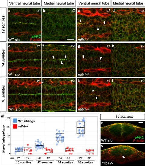
Notch signaling is required for the establishment of apico-basal polarity in the dorso-medial spinal cord. (a–l) Confocal sections taken at different dorso-ventral levels of the anterior spinal cord of WT sibling and mib1 mutant embryos. Dorsal views, anterior left. z1 corresponds to the ventral-most extent of apico-basally polarized neuro-epithelial tissue (identified by aPKC staining), z2 is localized 12 µm more dorsally in the same embryo. Arrowheads indicate local foci of polarized aPKC in partially polarized tissue. (a–d) At the 12 somites stage polarized aPKC signal is detected in the ventral-most neural tube in WT sibling and mib1 mutants. (e-l) At later stages polarity is progressively established in more dorsal regions of the neural tube in WT siblings (f,j), but remains limited to the ventral neural tube in mib1 mutants (g,h,k,l). (m) Quantification of the progressive emergence of apico-basally polarity in the anterior spinal cord (see Methods). Boxes represent mean values ± SD. (n,o) Transversal sections (dorsal up) through the neural tube of 14 somites stage embryos. (n) Polarized aPKC staining starts to spread through the dorso-ventral extent of the neural tube in WT siblings. (o) In mib1 mutants polarized aPKC enrichment remains limited to the ventral floor plate region (arrowhead). Scalebars: 20 µm.
|
