|
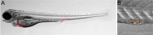
M. marinum infection of zebrafish embryos. (A) Fluorescence and bright-field overlay image of a zebrafish larva at 5 dpf showing the formation of granuloma-like cell aggregates containing red fluorescently labeled M. marinum bacteria. (B) Confocal microscopy and bright-field (DIC) overlay image of a granuloma-like aggregate in the tail of a zebrafish larva at 7 dpf. M. marinum bacteria in the aggregate show expression of a constitutive mCherry reporter and a granuloma-activated GFP reporter (GFP driven by the gag7 (granuloma-activated gene 7) promoter [63]. Embryos were infected by injection of M. marinum bacteria into the blood island at 1 dpf. Scale bar 25 µm.
|