FIGURE SUMMARY
Title

Host-Pathogen Interactions Made Transparent with the Zebrafish Model

Authors
Meijer, A.H., and Spaink, H.P.
Source
Full text @ Curr. Drug Targets

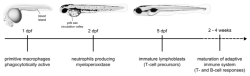
Schematic overview of the development of the zebrafish immune system. Commonly used sites for systemic bacterial infection of embryos by microinjection are the blood island and the yolk sac circulation valley at 1-3 dpf.

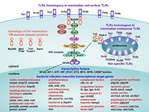
Components of the TLR pathway and genes commonly induced during the innate immune response of zebrafish to bacterial infection. Annotation of the zebrafish TLRs is based on Meijer et al. [118]. Cell surface or endosomal localizations of the zebrafish TLRs are hypothetical, based on localization of their mammalian homologs. The fish specific TLRs are tentatively placed on the endosome, since members of this group have been shown to recognize DNA or RNA ligands similar to the mammalian endosomal TLRs [169, 170]. The zebrafish genome encodes four TIR-domain adaptor proteins and lacks a homolog of the mammalian TRAM/Ticam2 adaptor [118-120]. Zebrafish Tlr5a/b and mammalian TLR5 share specificity for the recognition of bacterial flagellin [26]. The pathway from Tlr5a/b to NFκB is based on data from mammalian models [116] and has not been experimentally confirmed in zebrafish. The pathway from Tlr3 to NFκB is based on Sullivan et al. [119]. A selection of common transcriptionally-induced target genes is shown, based on S. typhimurium infection experiments in zebrafish embryos [26, 30] (unpublished results). Many of these genes are also induced in adult zebrafish at the end stage of M. marinum infection [25, 27, 28]. The indicated transcription factor families are induced by TLR signaling or by other pathways involved in the innate immune response. Several members of these transcription factor families as well as several components of the TLR pathway also show induced expression levels during bacterial infections [25-28].

M. marinum infection of zebrafish embryos. (A) Fluorescence and bright-field overlay image of a zebrafish larva at 5 dpf showing the formation of granuloma-like cell aggregates containing red fluorescently labeled M. marinum bacteria. (B) Confocal microscopy and bright-field (DIC) overlay image of a granuloma-like aggregate in the tail of a zebrafish larva at 7 dpf. M. marinum bacteria in the aggregate show expression of a constitutive mCherry reporter and a granuloma-activated GFP reporter (GFP driven by the gag7 (granuloma-activated gene 7) promoter [63]. Embryos were infected by injection of M. marinum bacteria into the blood island at 1 dpf. Scale bar 25 µm.

Acknowledgments
This image is the copyrighted work of the attributed author or publisher, and ZFIN has permission only to display this image to its users. Additional permissions should be obtained from the applicable author or publisher of the image. Full text @ Curr. Drug Targets