
Figure 4
- ID
- ZDB-FIG-190723-2018
- Publication
- Mehta et al., 2014 - The Cellular and Physiological Functions of the Lowe Syndrome Protein OCRL1
- Other Figures
- All Figure Page
- Back to All Figure Page
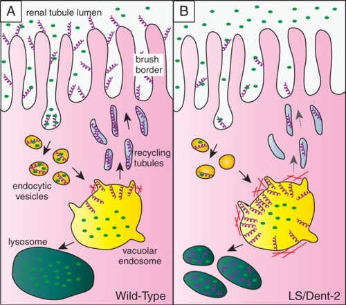
Model for OCRL1 function in endocytic trafficking of megalin in the renal tubule. A) The multiligand receptor megalin (purple helices) is abundant at the apical membrane of the epithelial cells lining the proximal tubule, where it binds to low-molecular-weight proteins in the renal filtrate (green ovals). Megalin is internalized by endocytosis and delivered via endocytic vesicles to the large vacuolar sorting endosomes found in this cell type. The low pH in the vacuolar endosome dissociates the megalin ligands, which in most cases are delivered to the lysosome for degradation. Megalin is sorted into recycling tubules that bud from the vacuolar endosome and deliver the receptor back to the plasma membrane for further rounds of endocytosis and recycling. B) Upon OCRL1 deficiency, megalin trafficking is impaired. Recycling of megalin from vacuolar early endosomes to the plasma membrane occurs less efficiently owing to aberrant accumulation of actin at the endosomal membrane. This results in endosomal accumulation of the receptor and missorting to the lysosome. The reduced abundance of megalin at the plasma membrane is responsible for reduced endocytosis of low-molecular-weight proteins from the renal filtrate, explaining the low-molecular-weight proteinuria seen in Lowe syndrome and Dent-2 disease. |