
- Title
-
The Cellular and Physiological Functions of the Lowe Syndrome Protein OCRL1
- Authors
- Mehta, Z.B., Pietka, G., and Lowe, M.
- Source
- Full text @ Traffic

Schematic diagram showing the organs affected in Lowe syndrome. Oculocerebrorenal syndrome of Lowe affects the eyes, central nervous system and kidneys, with specific manifestations in each organ as indicated. Dent-2 affects the same organs and displays similar manifestations, although the ocular and neurological defects are typically milder than those seen in Lowe syndrome. Renal tubular acidosis is also less frequently observed in Dent-2 disease. |

Cellular localization of OCRL1. A) OCRL1 (blue hexagons) has been localized to a number of cellular compartments. It is present at the TGN and various compartments of the endocytic pathway, where it resides at late-stage clathrin-coated pits, clathrin-coated vesicles, signaling endosomes, early or sorting endosomes and on recycling endosomes. OCRL1 has also been localized to the basal body of primary cilia, and it may also localize to the cilium itself. In maturing epithelia, OCRL1 transiently localizes to adherens and tight junctions. OCRL1 is recruited to phagosomes at a late stage in their formation, and is important for closure of the phagocytic cup as well as signaling events that occur post-sealing. OCRL1 is recruited to phagosomes generated by invading pathogenic bacteria such as |
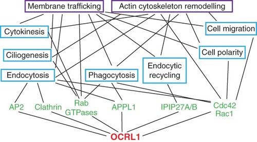
Network diagram showing the cellular functions of OCRL1. OCRL1 interaction partners are shown in green, and associated functions are indicated in blue boxes. Purple boxes highlight the two universal processes that are influenced by OCRL1, namely membrane trafficking and actin cytoskeleton remodeling. Both are relevant for all the functions shown in blue, and are linked accordingly. Note the diagram is not exhaustive, and some of the OCRL1-binding partners are likely to participate in several of the processes indicated. Although endocytosis and endocytic recycling are types of membrane trafficking, they are also shown separately as distinct processes given their reliance on different OCRL1-binding proteins. |
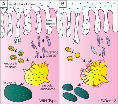
Model for OCRL1 function in endocytic trafficking of megalin in the renal tubule. A) The multiligand receptor megalin (purple helices) is abundant at the apical membrane of the epithelial cells lining the proximal tubule, where it binds to low-molecular-weight proteins in the renal filtrate (green ovals). Megalin is internalized by endocytosis and delivered via endocytic vesicles to the large vacuolar sorting endosomes found in this cell type. The low pH in the vacuolar endosome dissociates the megalin ligands, which in most cases are delivered to the lysosome for degradation. Megalin is sorted into recycling tubules that bud from the vacuolar endosome and deliver the receptor back to the plasma membrane for further rounds of endocytosis and recycling. B) Upon OCRL1 deficiency, megalin trafficking is impaired. Recycling of megalin from vacuolar early endosomes to the plasma membrane occurs less efficiently owing to aberrant accumulation of actin at the endosomal membrane. This results in endosomal accumulation of the receptor and missorting to the lysosome. The reduced abundance of megalin at the plasma membrane is responsible for reduced endocytosis of low-molecular-weight proteins from the renal filtrate, explaining the low-molecular-weight proteinuria seen in Lowe syndrome and Dent-2 disease. |