
Fig 6
- ID
- ZDB-FIG-190723-1386
- Publication
- Buchan et al., 2019 - A transgenic zebrafish line for in vivo visualisation of neutrophil myeloperoxidase
- Other Figures
- All Figure Page
- Back to All Figure Page
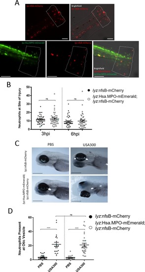
Transgene expression does not disrupt neutrophil recruitment to sites of injury or infection. A) Non-humanised (lyz:nfsB-mCherry only) and humanised (lyz:MPO-mEmerald; lyz:nfsB-mCherry) 3dpf larvae with tailfins transected to induce neutrophil recruitment; dashed outline represents the area in which neutrophils were counted. Scale bar = 250μm. B) Neutrophils present at the site of injury at 3 and 6 hours post injury (hpi); blue points denote the representative images in A). Error bars shown are mean ± SEM (n = 45 over three independent experiments); groups were analysed using an ordinary two-way ANOVA and adjusted using Bonferroni’s multiple comparisons test; ns, p>0.9999. C) Non-humanised and humanised larvae injected with either a PBS vehicle control or 1,400cfu S. aureus USA300 into the otic vesicle at 3dpf, then fixed in paraformaldehyde at 4 hours post infection (hpi) and stained with Sudan Black B to detect neutrophils; dashed white outline indicates the otic vesicle. D) Neutrophils present at the otic vesicle at 4hpi. Scale bars = 250μm. Error bars shown are mean ± SEM (n = 25 over two independent experiments); groups were analysed using an ordinary two-way ANOVA and adjusted using Bonferroni’s multiple comparisons test. ****, p<0.0001; ns, p>0.9999. |