
Fig. 3
- ID
- ZDB-FIG-190718-10
- Publication
- Eno et al., 2019 - Aggregation, segregation and dispersal of homotypic germ plasm RNPs in the early zebrafish embryo
- Other Figures
- All Figure Page
- Back to All Figure Page
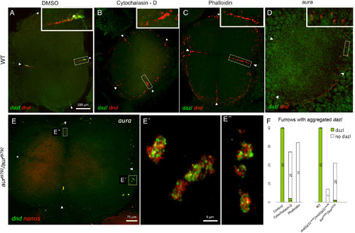
originally localized to the vegetal cortex, accumulates at the furrows (arrowheads) of the four cell‐staged blastocyst at the animal pole in wild‐type control (DMSO‐treated) embryos (A) but not in wild‐type embryos treated with F‐actin drugs cytochalasin‐D (B) and phalloidin (C), yet drug‐treated embryos exhibit germ plasm aggregate formation. D–E″:aura/mid1ip1Lmutant embryos show severely reduced dazl RNP accumulation at the furrow (Welch and Pelegri, 2015) (D), yet contain animal RNP aggregates (dnd/nanos, E, E″). F: Quantitation dnd RNP aggregates in the presence or absence of dazlRNPs: Panels A–E = confocal Z‐projections; Panels E′–E″ = SIM Z‐projections. Scale bars = 100 μm in A–D; 75 μm in E; 8 μm in E′–E″. |