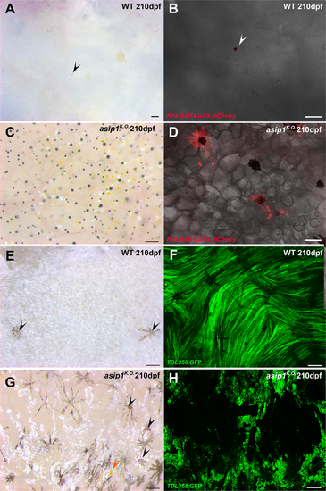
Detailed visualization of ventral pigment cells in WT and asip1 mutants. (A) Ventral view of 210 dpf WT belly. (B) Belly of 210 dpf WT fish carrying Tg(Kita:GalTA4;UAS:mCherry) (labels melanophores) transgene shows no melanophores in ventral skin. (C) Ventral view of 210 dpf asip1K.O. belly. (D) Belly of 210 dpf asip1K.O. fish carrying Tg(Kita:GalTA4;UAS:mCherry) transgene shows high number of melanophores in ventral skin. (E) Internal view of 210 dpf WT abdominal wall shows a white sheet of iridophores with few internal melanophores (black arrow). (F) Abdominal wall of 210 dpf WT fish carrying Tg(TDL358:GFP) (labels iridophores and glia) transgene displays a uniform and continuous sheet of iridophores. (G) Internal view of 210 dpf asip1K.O. abdominal wall shows a disrupted and discontinuous sheet of iridophores with high number of melanophores (black arrow) and some xanthophores (orange arrow). (H) Abdominal wall of 210 dpf asip1K.O. fish carrying Tg(TDL358:GFP)transgene exhibits a broken sheet of iridophores. Scale bars: 100 μm.
|
