
Fig. 7
- ID
- ZDB-FIG-190627-31
- Publication
- Campo-Paysaa et al., 2019 - Generation of the squamous epithelial roof of the 4th ventricle
- Other Figures
- All Figure Page
- Back to All Figure Page
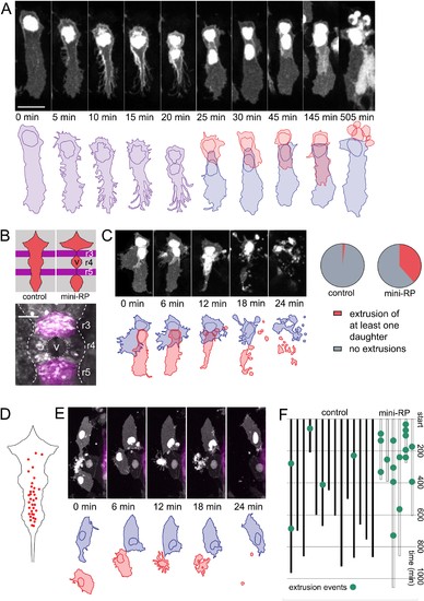
Space-regulated cell extrusions in the roof plate.(A) Time-lapse frames of veil cell division starting at 24 hpf with cleavage plane close to parallel to roof plate border generates daughters with same fates (two veil cells). The lateral daughter (blue in schematic) inherits the basolateral veil while the medial daughter (red in schematic) attempts to regenerate a new veil but becomes extruded. A schematic representation of the sequence is shown in the bottom panel. (B) Schematic of the dorsal hindbrain in control and mini-RP (Krox20 x Rab11dn) embryos, showing the failure in ventricle opening at the level of rhombomeres 3 and 5 and consequent restriction of ventricle and roof plate in rhombomere four in the latter. Photomicrograph is maximum projection of rhombomeres 3, 4 and 5 in a mini-RP embryo showing mini-ventricle in rhombomere four and lack of ventricles in rhombomeres 3 and 5 (magenta). (C) Time-lapse sequence of the extrusions of two sister veil cells in the roof plate of rhombomere four in a mini-RP embryo. A schematic of the sequence is shown below. Pie charts show the quantification of the proportion of veil cell divisions producing extruding daughter cells, in control (n = 21) and mini-RP embryos (n = 13). (D) Schematic shows map of 35 extrusions of squamous roof plate cells in control embryos. (E) Time-lapse sequence of a squamous cell (red cell in schematic) being extruded from roof plate of rhombomere 2, in a mini-RP embryo. Top cell in images (and blue in schematic) is a veil cell lying over rhombic lip. (F) Temporal maps of squamous cell extrusions in the roof plate of rhombomere 4, in control (black lines) and mini-RP (white lines) embryos. Each vertical line represents a log of cell extrusion events during a single time-lapse recording. Scale bars: 10 μm in A. |