
- Title
-
Generation of the squamous epithelial roof of the 4th ventricle
- Authors
- Campo-Paysaa, F., Clarke, J.D., Wingate, R.J.
- Source
- Full text @ Elife
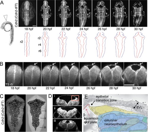
(A) Left. Diagram of 24 hpf zebrafish in lateral view. Arrowhead indicates location of hindbrain roof plate covering fourth ventricle and angle of imaging for time-lapse sequence. Right. Selected frames from time-lapse of Cdh2:(Cdh2-tFT) embryo (n = 15) tracking opening and expansion of fourth ventricle from 18 hpf to 32 hpf (see also Figure 1—video 1). White arrowheads on photomicrographs indicate the three points at which widening commences. Cdh2 is ubiquitous in neural cell membranes and most highly expressed at the ventricular surface. Brightest expression reveals the location of the rhombic lips which sit at interface between columnar neuroepithelium and the periphery of squamous roof plate. Outlines of the neural tube (black) and rhombic lip (red) are shown below. Roof plate widening is initiated in even-numbered rhombomeres (r), 2, 4, 6. (B) Outfolding of dorsal columnar epithelium and de novo appearance of the squamous roof plate (arrows) is revealed in transverse plane time-lapse of ventricle opening at level of rhombomere 5 (Figure 1—video 2). (C) Maximum projection of confocal sections of squamous roof plate and attached columnar epithelium in dorsal view of hindbrain in a 24 hpf Cdh2:(Cdh2-tFT) embryo. Columnar epithelium has been greyed out in right hand image to reveal shape of squamous roof plate. (D) Transverse plane reconstructions through levels indicated in (C) show shape of ventricle and domed squamous roof plate at different anteroposterior levels. (E) Electron micrograph of region indicated by inset in (D) of epithelial transition zone between squamous roof plate (pink) and columnar neuroepithelium of rhombic lip (blue and green). The most dorsal neuroepithelial cell, which abuts the roof plate at the transition zone between tissue architectures, is highlighted in yellow. Images in A-D are from Cdh2:(Cdh2-tFT) embryos. EVL, enveloping layer. Scale bars: 50 μm in A and B. |

(A) Prior to ventricle expansion cells at dorsal midline of the neural tube are irregular and overlap the midline, while slightly deeper columnar cells align their apical end feet to the midline. (B) Pard3-GFP (yellow) is unpolarised in irregular dorsal cells but already polarized to the apical pole of columnar neuroepithelial cells that lie just deep to the dorsal surface of the neural tube. Transverse plane view of neural tube indicates level of sections in (A) and (B, C) Schematic diagrams of neural tubes show location of cells illustrated in panels (D) to (G, D) Three frames from time-lapse starting at 18hpf show two superficial cells at the dorsal midline (dashed line in drawings) initially overlap but later share a contiguous border that maps to the geometric midline just prior to opening of the ventricle. (E) Three frames from time-lapse show two spikey midline cells at dorsalmost aspect of neural tube transform directly into squamous roof plate cells as the ventricle opens over 4 hr. Dotted lines in drawings represent borders of roof plate (see Figure 2—video 1). (F) Four frames from time-lapse starting at 21hpf show irregular dorsal midline cell (nucleus marked by yellow dot on left hand images and cytoplasm filled yellow in adjacent right hand images) migrating along the dorsal tube midline to seed squamous roof plate as ventricle opens. A second roof plate pioneer is marked with blue dot and blue cytoplasm. Dotted lines in images show borders of roof plate and lateral edges of neural tube (see Figure 2—video 2). (G) Pard3-GFP protein is progressively allocated to cell-cell junctions surrounding a squamous roof plate cell during the opening of the ventricle. Scale bar in (G): 20 μm. |
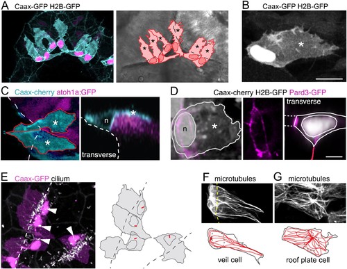
A cell with novel morphology, the ‘veil’ cell, occupies the transition zone between squamous roof plate and columnar neuroepithelium.(A) Dorsal view of six cells in the transition zone in the rostral pole of the hindbrain roof plate. Cells have distinctive morphologies with irregular lateral veils (asterisked in schematic to right) spanning the interface (dotted line) between roof plate and the columnar neuroepithelium. (B) Single veil cell with a nucleus placed at periphery of roof plate territory and a lateral veil (asterisk) extending over neuroepithelium. (C) Left. A dorsal view of two veil cells (cyan) with lateral veils (asterisks) extending over rhombic lip progenitors expressing atoh1a:GFP (magenta). Roof plate border shown with dashed line. Right. In transverse view reconstruction a veil cell presents a characteristically comma-shaped profile as its veil (asterisk) wraps itself over the basolateral surface of the atoh1-positive neural rhombic lip precursor. Apical surface of ventricle shown dashed, nucleus indicated with n. (D) Left: Dorsal view of veil cell, its nucleus (n) and veil (asterisk). Pard3-GFP expression shown in magenta. Middle: Single channel showing this cell’s apical ring of Pard3-GFP. Right: Same cell reconstructed in transverse plane showing Pard3-GFP (magenta) expression at the ventricular interface with squamous epithelium (dashed lines) and apical surface of columnar epithelium (red line). (E) Cilia (white) of veil cells (magenta) are located close to nuclei and protrude into ventricle. Roof plate border shown dashed. Many cilia from columnar cells are also visible in the maximum projection. (F) Two adjacent veil cells expressing Dck1k-GFP show highly polarized microtubule networks, comprising a near linear array extending into the veils. (G) By comparison, central cell in this image is a squamous roof plate cell displaying radially organized microtubule array around a centrally placed focal point. |
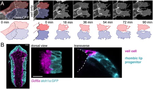
Veil cells comprise a morphologically dynamic, contiguous gdf6a-positive population that wraps over the rhombic lip.(A) Dorsal view of time-lapse sequence of two adjacent veil cells highlighting dynamics of their basolateral veils (blue and red, see also Figure 4—video 1). Outline drawings of veil dynamics shown below. (B) Left. Dorsal view of hindbrain showing in situ hybridisation for Gdf6a (magenta) and atoh1a-GFP rhombic lip progenitors (cyan). Right. High magnification of inset area in (g) and a transverse plane reconstruction. |

Veil cells can transform into definitive squamous roof plate cells.(A) Four frames from a time-lapse of caax-RFP labeled veil cell starting at 22 hpf transforming directly to a roof plate cell. Retraction of the veil from columnar epithelium is indicated with arrowheads. Dashed red line is boundary between columnar epithelium to its left and squamous epithelium to its right (see Figure 5—video 1). (B) Schematic shows map of 16 veil cells that transformed directly to roof plate cells, most lie in the lower rhombic lip. |
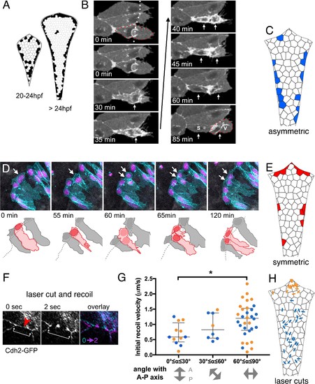
(A) Schematics of roof plate that cumulatively map the distribution of cell divisions between 20 and 24 hpf and from 24 to 36 hpf from 125 embryos. (B) Time-lapse sequence of veil cell (outlined by red dots) during an asymmetric division (see also Figure 6—video 1) with cleavage plane parallel to roof plate border (dotted white line). Two timepoints in mitosis indicated by single white arrow at 30 and 35 min. Position of two daughters indicated by white arrows from 40 min onwards. Medial daughter becomes a squamous roof plate cell (marked S) and lateral daughter becomes a veil cell (marked V and outlined by red dots). Images also contain a second veil cell and an established squamous cell that have been indicated with reduced brightness in the drawing. (C) Schematic map of location of 26 asymmetrically fated veil cell divisions. (D) Five frames from time-lapse of a symmetrically fated veil cell division starting at 23 hpf that generates daughters with same fates (two veil cells). Arrows indicate mother and daughter veil cells. One daughter inherits the original basolateral veil while the other rapidly regenerates a veil after mitosis (see also Figure 6—video 2). (E) Schematic map of 19 symmetrically fated veil cell divisions. (F) Time-lapse sequence (see also Figure 6—video 3) showing laser cut of a cell-cell junction in the roof plate of a Cdh2-GFP embryo at 24hpf. Site of laser ablation is indicted with a red arrowhead. Arrows indicate positions and recoil of adjacent tricellular vertices. (G) Quantification of initial recoil velocity after laser cut as a function of the angle to the anteroposterior axis shows a significantly higher velocity following parallel versus perpendicular cuts (Two-tailed Mann-Whitney, p=0.0113). Recoil in the region proximal to the upper rhombic lip (orange) is similar to that adjacent to the lower rhombic lip (blue). (H) Map of positions and angles of laser cuts. |
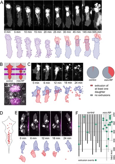
Space-regulated cell extrusions in the roof plate.(A) Time-lapse frames of veil cell division starting at 24 hpf with cleavage plane close to parallel to roof plate border generates daughters with same fates (two veil cells). The lateral daughter (blue in schematic) inherits the basolateral veil while the medial daughter (red in schematic) attempts to regenerate a new veil but becomes extruded. A schematic representation of the sequence is shown in the bottom panel. (B) Schematic of the dorsal hindbrain in control and mini-RP (Krox20 x Rab11dn) embryos, showing the failure in ventricle opening at the level of rhombomeres 3 and 5 and consequent restriction of ventricle and roof plate in rhombomere four in the latter. Photomicrograph is maximum projection of rhombomeres 3, 4 and 5 in a mini-RP embryo showing mini-ventricle in rhombomere four and lack of ventricles in rhombomeres 3 and 5 (magenta). (C) Time-lapse sequence of the extrusions of two sister veil cells in the roof plate of rhombomere four in a mini-RP embryo. A schematic of the sequence is shown below. Pie charts show the quantification of the proportion of veil cell divisions producing extruding daughter cells, in control (n = 21) and mini-RP embryos (n = 13). (D) Schematic shows map of 35 extrusions of squamous roof plate cells in control embryos. (E) Time-lapse sequence of a squamous cell (red cell in schematic) being extruded from roof plate of rhombomere 2, in a mini-RP embryo. Top cell in images (and blue in schematic) is a veil cell lying over rhombic lip. (F) Temporal maps of squamous cell extrusions in the roof plate of rhombomere 4, in control (black lines) and mini-RP (white lines) embryos. Each vertical line represents a log of cell extrusion events during a single time-lapse recording. Scale bars: 10 μm in A. |