
Fig. 3
- ID
- ZDB-FIG-190626-50
- Publication
- Chopra et al., 2019 - Zebrafish duox mutations provide a model for human congenital hypothyroidism
- Other Figures
- All Figure Page
- Back to All Figure Page
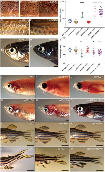
Adult duox mutant zebrafish display an array of visible phenotypes. (A–C) 5× magnification of flank region showing the distribution of melanophores in WT, sa9892+/− and sa989−/− siblings. The apparent abundance of melanophores was statistically significant in duoxmutants (D). Asterisks denote statistically significant differences (Bonferroni's multiple comparisons test, ****P<0.0001). duox mutants also showed irregularities in stripe pattern in contrast to heterozygous siblings, shown here in a 2× magnification of the flank in sa9892siblings (E,F). Craniofacial anomalies were evident among mutants, with frontal height significantly shorter among mutants (G–I) (Bonferroni's multiple comparisons test, *P<0.5, **P<0.01). Erythema in the thoracic region was prominent among mutants. This was especially noticeable in nacre backgrounds (L,N,O). duox mutants also suffered from perpetual fin damage, which manifest as ragged margins and tears (S–U). Scale bars: 1 mm. |
| Fish: | |
|---|---|
| Observed In: | |
| Stage: | Adult |