
Fig. 4
- ID
- ZDB-FIG-190626-41
- Publication
- Perez et al., 2019 - Mutations in the microtubule-associated protein MAP11 (C7orf43) cause microcephaly in humans and zebrafish
- Other Figures
- All Figure Page
- Back to All Figure Page
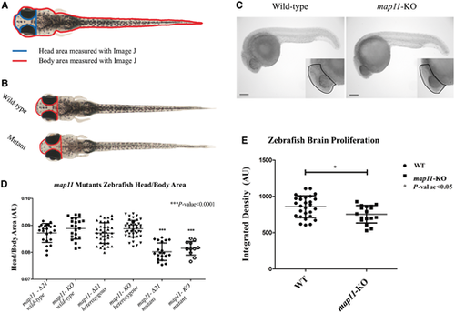
CRISPR/Cas9-mediated MAP11-orthologue zebrafish mutants recapitulate the human microcephaly phenotype. (A) Head to body area measurements of CRISPR/Cas9 MAP11 orthologue mutants. Zebrafish head and body surface area were measured at age 28 days post fertilization using ImageJ software. Head to body area ratios are plotted. (B) Wild-type and homozygous map11-Δ21 mutant zebrafish (28 days post fertilization) demonstrating same body area surface. A red line highlights the head surface area of each larva showing smaller head of map11-Δ21 mutants compared to wild-types. (C) Images of 24 hpf zebrafish embryos stained for phospho-Histone H3. Images of map11-KO embryos show decreased proliferating cells within brain regions compared to wild-types (black dots). Scale bar = 0.5 mm. (D) Head-to-body ratios of map11-KO and map11-Δ21homozygous mutants. P-values represent comparison of each mutant (map11-Δ21 and map11-KO) to its corresponding wild-type control. P-values were calculated using an unpaired two-sided student t-test. (E) A graph showing greater proliferation capacity of wild-type (n = 28) compared to map11- KO (n = 17) zebrafish brains (measured by integrated density of the region of interest). |
| Fish: | |
|---|---|
| Observed In: | |
| Stage Range: | Prim-5 to Days 21-29 |