
Fig. 4
- ID
- ZDB-FIG-190626-13
- Publication
- Gerber et al., 2019 - The HMG box transcription factors Sox1a and b specify a new class of glycinergic interneurons in the spinal cord of zebrafish embryos
- Other Figures
- All Figure Page
- Back to All Figure Page
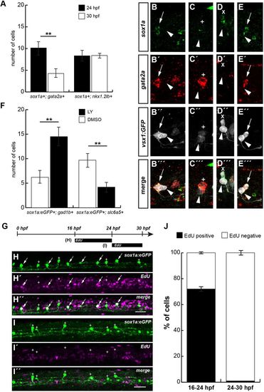
V2s neurons depend on Notch signalling. (A) Mean number of cells expressing sox1a and gata2a mRNA at 24 hpf (black) and 30 hpf (white) determined by FISH (Fig. S5A-B″). Cells expressing sox1a and gata2a mRNA decreased by about 48% from 24 (black, n=71) to 30 hpf (white, n=34), whereas sox1a:eGFP+ V2 cells co-expressed nkx1.2lb mRNA (Fig. S5C-D″) at similar levels at both stages. (B-E‴) Examples of co-expression of vsx1:GFP, gata2a and sox1amRNA. (B-B‴) A pair of vsx1:GFP+ cells with one cell being vsx1:GFP+;gata2a+;sox1a+ (arrows) extending axons at 24 hpf. Committed V2a cells express only vsx1:GFP (arrowheads). (C-C‴) Example of a vsx1:GFP+ cell co-expressing gata2a (+) at 24 hpf. (D-D‴) A V2s cell that is still vsx1:GFP+ and expresses sox1a mRNA (x) at 26 hpf. (E-E‴) sox1a is co-expressed with gata2a in one cell of a V2a/V2b,s pair (arrows) at 22 hpf. Arrowheads indicate vsx1:GFP+ V2a neurons. (F) Disruption of Notch signalling in sox1a:eGFP embryos from 16 to 24 hpf and assessment of neurotransmitter type at 30 hpf shows a 2.3-fold increase in sox1a:eGFP+ neurons expressing the GABAergic marker gad1b. In contrast, blocking of Notch signalling leads to a 2.3-fold decrease in sox1a:eGFP+;slc6a5+ neurons (DMSO-treated control, white; LY treated, black; for original data, see Fig. S5E-H″). (G-J) sox1a precursor cells divide largely before 24 hpf. sox1a:eGFP+ (green) embryos were treated with EdU (magenta) during two different time windows before processing with EdU click-chemistry at 30 hpf (G). sox1a:eGFP embryos treated from 16 to 24 hpf (H-H″) or from 24 to 30 hpf (I-I″) showing a five-somite spinal cord segment at 30 hpf (sox1a:eGFP+; EdU+ cells, arrows; sox1a:eGFP+; EdU− cells, +). (J) Percentages of EdU+and EdU−; sox1a:eGFP+ cells. Most cells divided before 24 hpf. (A,F) Counts of six to nine embryos from two independent experiments in the V2 domain of the spinal cord above the yolk extension over a five-somite distance. Dorsal is upwards; anterior is leftwards. Data are mean±s.e.m. (A,F) or mean±s.d. (J). Statistical significance was assessed using the unpaired two-tailed Student's t-test. **P≤0.01. Scale bars: 25 µm. |
| Genes: | |
|---|---|
| Fish: | |
| Anatomical Terms: | |
| Stage: | Prim-5 |