
Fig. 3
- ID
- ZDB-FIG-190626-11
- Publication
- Gerber et al., 2019 - The HMG box transcription factors Sox1a and b specify a new class of glycinergic interneurons in the spinal cord of zebrafish embryos
- Other Figures
- All Figure Page
- Back to All Figure Page
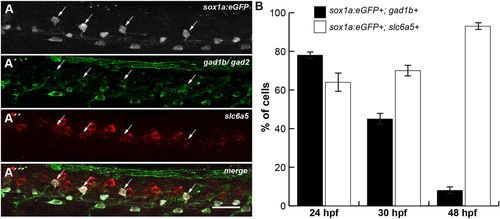
V2s cells develop into glycinergic interneurons. (A-A‴) Multicolour-labelling of a 24 hpf sox1a:eGFP transgenic embryo using FISH with a slc6a5 probe (red), and immunohistochemistry (IHC) with anti-eGFP (white) and anti-Gad1b/Gad2 (green) antibodies. Many sox1a:eGFP+ cells in the V2 domain are both GABA- and glycinergic at 24 hpf (arrows). (B) Percentage of cells expressing sox1a:eGFP and gad1b (black) or sox1a:eGFP and slc6a5 (white) calculated from FISH with a gad1b or slc6a5 probe, and IHC with anti-eGFP antibody at 24, 30 and 48 hpf (Fig. S4). At 24 hpf, 78% of sox1a:eGFP+ cells are GABAergic (n=91 of 117) and 64% are glycinergic (n=34 of 53). At 30 hpf, 45% of sox1a:eGFP+ cells (n=51 of 114) are GABAergic and 70% are glycinergic (n=56 of 80). At 48 hpf, only a minor fraction (8%) still expresses gad1b (n=8 of 98). The majority of sox1a:eGFP cells (93%) have turned on the glycinergic marker by 48 hpf (n=84 of 90). Counts of five to eight embryos from two independent experiments. Data are mean±s.d. Views of spinal cord over yolk extension: dorsal is upwards; anterior is leftwards. Scale bar: 25 µm. |
| Gene: | |
|---|---|
| Antibody: | |
| Fish: | |
| Anatomical Terms: | |
| Stage Range: | Prim-5 to Long-pec |