
Fig. 5
- ID
- ZDB-FIG-190624-18
- Publication
- Obermann et al., 2019 - The Surface Proteome of Adult Neural Stem Cells in Zebrafish Unveils Long-Range Cell-Cell Connections and Age-Related Changes in Responsiveness to IGF
- Other Figures
- All Figure Page
- Back to All Figure Page
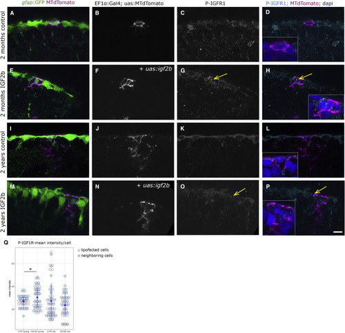
IGF2b Overexpression Activates the IGF1R in Lipofected Cells (A–P) P-IGF1R immunohistochemistry on young (A–H) and old (I–P) brains. The radial glia are labeled by the gfap:GFP transgene. Single cells in magenta have been lipofected in vivo 4 days prior to brain fixation with mtdTomato (control, A–D and I–L) or with mtdTomato and igf2b (IGF2b, E–H and M–P), highlighting primarily the cell soma (the radial process is not always in the plane of the optical section, but it is present below the somata of lipofected cells). We observe the distinct expression levels of P-IGF1R in young control (C and D), young + IGF2b (highest, arrows in G and H), old control (K and L), and old + IGF2b (a few dots, arrows in O and P). Insets in (D), (H), (L), and (P) are close-ups of the lipofected cells; nuclei in blue stained by DAPI. Single confocal planes are displayed. Scale bar, 10 μm. (Q) The mean intensity of the phospho-IGF staining was measured on 7 lipofected cells and their immediate neighbors in each condition. Staining intensities differ significantly between control and igf2b-lipofected brains (t test; p = 0.030) in young but not in old brains. |