
FIGURE
Fig. 7
- ID
- ZDB-FIG-190530-18
- Publication
- Konjikusic et al., 2018 - Mutations in Kinesin family member 6 reveal specific role in ependymal cell ciliogenesis and human neurological development
- Other Figures
- All Figure Page
- Back to All Figure Page
Fig. 7
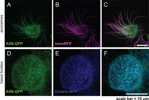
Kif6-GFP localizes to the basal bodies and axonemes of Xenopus multi-ciliated cells. Confocal imaging of the mucocilated Xenopus laevis epidermis demonstrating consistent Kif6-GFP localization within the axonemes (green; A, C) and at the basal bodies (green; D, F). Expression of pan-membrane-RFP marker (magenta; B, C) to co-label the axonemes and Centrin-BFP (blue; E, F) to co-label the basal bodies. Scale Bars: 10μm. |
Expression Data
Expression Detail
Antibody Labeling
Phenotype Data
Phenotype Detail
Acknowledgments
This image is the copyrighted work of the attributed author or publisher, and
ZFIN has permission only to display this image to its users.
Additional permissions should be obtained from the applicable author or publisher of the image.
Full text @ PLoS Genet.