
Fig. 5
- ID
- ZDB-FIG-190530-16
- Publication
- Konjikusic et al., 2018 - Mutations in Kinesin family member 6 reveal specific role in ependymal cell ciliogenesis and human neurological development
- Other Figures
- All Figure Page
- Back to All Figure Page
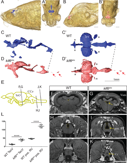
(A-B’) 3D-reconstruction of representative iodine-contrasted μCT dataset from WT (A-A’) and kif6sko mutant zebrafish at 90dpf. (C-D’) 3D-reconstruction and segmentation of virtual endocasting of ventricular system in WT (blue; C-C’) and kif6sko mutant (red; D-D’) zebrafish from datasets in A-B’ demonstrated morphological alterations of the ventricular system including dilation of the central canal (red arrows; D-D’) and stenosis of small ventricles (asterisks, D-D’). (E) Schematic of adult zebrafish brain highlighting the relative transverse optical section of the zebrafish brain in WT (F, H, J) and kif6sko homozygous mutant (G, I, K) zebrafish brain at 90dpf. (F, G) The medial region of the TeO showing the medial TecV (yellow dashed line) which is dilated in kif6sko mutant fish (G) compared to age-matched WT (F). (H, I) Sectioning at the region of the medulla oblongata posterior to the lobus facialis showing dysmorphogenesis and deepening of the posterior RV (yellow dashed line) in kif6sko mutants (I) compared with WT (H) zebrafish. (J, K) Spinal cord sectioning showing dilation of the central canal in kif6sko mutant (K) compared to WT (J) zebrafish. (L) Quantitation of the areas (yellow dashed line) of the TecV and the RV posterior to the lobus facialis (pos. RV) in WT and kif6sko mutant zebrafish, highlighting a consistent dilation in kif6sko mutants (n = 11 sections/genotype; two-tailed t-test; ****, p<0.0001). Scale Bars: 1mm. DiV—diencephalic ventricle; TecV-tectal ventricle TeO-tectum opticum; CCe-corpus cerebelli; RV- rhombencephalic ventricle; Vam—medial division of valvula cerebelli; and Cc-central canal. |
| Fish: | |
|---|---|
| Observed In: | |
| Stage: | Adult |