
Fig. 2
- ID
- ZDB-FIG-190523-3
- Publication
- Lalonde et al., 2018 - Contributions of 5'HoxA/D regulation to actinodin evolution and the fin-to-limb transition
- Other Figures
- All Figure Page
- Back to All Figure Page
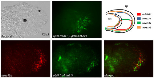
The“m-Inta11” regulatory element is active in a subpopulation of hoxd13a- and hoxa13a-expressing cells in the zebrafish pectoral fin at 72hpf. (A, D-F) Double fluorescent ISH for hoxa13a, eGFP, and (B) Tg(m-Inta11:eGFP) reporter activity in 72hpf pectoral fin. (C) Summary of expression patterns for hoxa13a, hoxd13a,hoxa13b and m-Inta11 activity in 72hpf pectoral fin. The “m-Inta11” regulatory element drives expression in the posterior fin fold mesenchyme (red arrow): eGFP fluoresence (B) and eGFP transcripts are presented (E). The expression of hoxa13a extends to the anterior fin fold mesenchyme (white arrow) (D,F), outside the region where “m-Inta11” is active (red arrow) (E,F). The expression of hoxd13a is posteriorly restricted and partially mimics “m-Inta11” activity (C). The “m-Inta11” element is not active in the proximal-posterior endoskeletal disc and fin fold regions where hoxd13a, and hoxa13b are co-expressed (purple arrow) (C). Brightfield (A), fluorescent (B,D,E) and merged (F) images are present. ED, endoskeletal disc; FF, fin fold. Scale bar, 3 mm. |