
Fig. 1
- ID
- ZDB-FIG-190328-9
- Publication
- Boyle Anderson et al., 2018 - A transcriptomics analysis of the Tbx5 paralogues in zebrafish
- Other Figures
- All Figure Page
- Back to All Figure Page
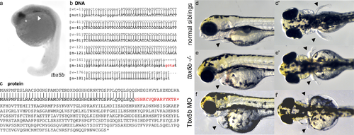
Tbx5b mutants phenocopy Tbx5b morphants. (a) tbx5b expression in the eyes (asterisk) and lateral plate mesoderm (arrow) of 21 hpf embryos. (b) The indel for the tbx5b-2A mutant (red) embryos is located in the beginning of the first intron. Uppercase letters are the protein coding sequence while lowercase letters are the untranslated regions and introns. (c) The mutation would produce a prematurely truncated protein with a frameshift resulting in incorrect amino acids starting at AA53 and a premature stop codon after 16 incorrect amino acids (red). (d-e) Embryos at 3 dpf. Arrows point to the heart and pectoral fins. Siblings from a Tbx5b in-cross display normal heart (d) and fin (d’) development while mutant siblings have affected hearts (e) and fins (e’) which phenocopy the defects seen in the heart (f) and fins (f’) of embryos injected with a Tbx5b morpholino. |
| Gene: | |
|---|---|
| Fish: | |
| Anatomical Terms: | |
| Stage: | 20-25 somites |
| Fish: | |
|---|---|
| Knockdown Reagent: | |
| Observed In: | |
| Stage: | Protruding-mouth |